
The new MRI technique developed by Hebrew University of Jerusalem researchers is especially important for understanding whether a patient is merely getting older or is developing a disease such as Parkinson’s or Alzheimer’s.
By Naama Barak
MRI, or magnetic resonance imaging, already does pretty amazing work giving us a better picture of what’s happening on our insides. But it is about to do even more amazing work, helping doctors determine more quickly the onset of neurodegenerative diseases.
A team of researchers from the Hebrew University of Jerusalem, led by Dr. Aviv Mezer, found a way for MRIs to record changes in the biological makeup of brain tissue. Their findings were published in Nature Communications.

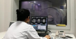
Patient being positioned for MRI study of the head. – Illustrative Photo: Ptrump16, Wikimedia
“Instead of images, our quantitative MRI model provides molecular information about the brain tissue we’re studying. This could allow doctors to compare brain scans taken over time from the same patient, and to differentiate between healthy and diseased brain tissue, without resorting to invasive or dangerous procedures such as brain tissue biopsies,” Mezer explained.
This ability is especially important for understanding whether a patient is merely getting older or is developing a disease such as Alzheimer’s or Parkinson’s.

Dr. Aviv Mezer of Hebrew University. Photo: courtesy
Both normal aging and neurodegenerative diseases create biological “footprints” in the brain, changing the lipid and protein content of brain tissue. Whereas existing MRI scans provide only pictures of the human brain, the new technique provides biological readouts of brain tissue—the ability to see what’s happening at a molecular level, and to direct a course of treatment accordingly.
This means patients will more likely receive correct diagnoses earlier, speeding up the beginning of their treatment and offering them an improved quality of life.
Looking forward, Mezer believes that the new MRI technique will also provide a crucial understanding into how our brains age.
“When we scanned young and old patients’ brains, we saw that different brain areas ages differently. For example, in some white-matter areas, there is a decrease in brain tissue volume, whereas in the gray-matter, tissue volume remains constant. However, we saw major changes in the molecular makeup of the gray matter in younger versus older subjects,” he said.
View original ISRAEL21c publication at:
https://www.israel21c.org/israeli-mri-method-could-enable-early-alzheimer-diagnosis/
‘as a light unto the nations’


 Israeli New Shekel Exchange Rate